Summer 2023 - Volume 59, Issue 2
Issue Overview
Show Filters
Figure 1. Hematite inclusions extend from the center of an aquamarine sample from Xinjiang, China. Photomicrograph by Yubing Chen; field of view 3.64 mm.
Internal World of Aquamarine from Xinjiang

Various inclusions are observed in a batch of aquamarine samples from Xinjiang, China.

Read More

Figure 1. This quartz crystal cluster from Larimer County, Colorado, contains numerous bluish green and white spherical inclusions on phantom growth planes. Photo by Annie Haynes; courtesy of Nico Jackson.
Clinochlore and Muscovite in Quartz from Colorado

Bluish green and white spheres create a beautiful inclusion scene in quartz from Larimer County, Colorado.

Read More

A subsurface feather in diamond resembling an insect wing, with visible iridescence caused by thin-film interference. Photomicrograph by Matthew Hardman; field of view 2.90 mm.
An Iridescent “Insect Wing” in Diamond

A feather inclusion with iridescence caused by thin-film interference in diamond resembles an insect wing.

Read More
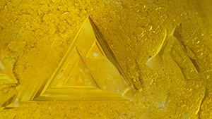
Trigons creating the impression of a thin and patchy “yellow skin” on a 0.98 ct rough diamond. Photomicrograph by Nicole Ahline; field of view 2.34 mm.
Patchy Yellow Trigon

Isolated nitrogen confined to the outer surface of a 0.98 ct diamond causes “yellow skin.”

Read More

These iridescence features observed in a 1.71 ct natural diamond (top) were reminiscent of Rainbow Mountain in Peru (bottom). Photomicrograph by Tejas Jhaveri; field of view 2.90 mm. Bottom photo courtesy of Daniel Sánchez Ibarra (Wikimedia Commons).
Rainbow Mountain in Natural Diamond

An iridescent feather inclusion in natural diamond bears resemblance to Rainbow Mountain in Peru.

Read More

A crystal with a stress halo and a smaller adjacent crystal resembles a dotted quarter note. Several reflections of such crystals with stress fractures are visible. Photomicrograph by Michaela Damba; field of view 2.90 mm.
Musical Diamond

A dark crystal and a stress cleavage crack resemble a quarter note in a diamond.

Read More
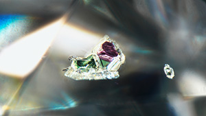
This “snail” inclusion was viewed through the upper half facet of a 4.00 ct near-colorless diamond. The purple-red “shell” was identified as pyrope garnet, while the green “body” was determined to be diopside. Photomicrograph by Christopher Vendrell; field of view 1.99 mm.
“Snail” in Diamond

A combination of pyrope garnet and diopside inclusions is reminiscent of a snail in a near-colorless diamond.

Read More

An opaque crystal, likely a metal sulfide, is suspended in a garnet. A combination of darkfield and oblique fiber-optic illumination makes it appear as though the crystal is hovering inside the stone. Portions of the inclusion are duplicated because of its split across several facet junctions, adding to the visual interest. Photomicrograph by E. Billie Hughes; field of view 4 mm.
Guest in Garnet: Metal Sulfide?

An intriguing metallic inclusion is observed in a faceted garnet.

Read More

This pristine hexagonal platelet of graphite was seen in a pink sapphire from Sri Lanka. Photomicrograph by Jeffrey Hernandez; field of view 1.99 mm.
Pink Sapphire with Graphite Inclusion

A hexagonal platelet of graphite is discovered in a pink sapphire from Sri Lanka.

Read More

This scene of an iron stain and numerous curved lines showing rainbow colors in a fracture resembles a scene from the night sky viewed through a telescope rather than a microscope. Photomicrograph by Charuwan Khowpong; field of view 2.85 mm.
Night Sky in Yellow Sapphire

Fiber-optic light creates a night sky scene in a yellow sapphire from Madagascar.

Read More